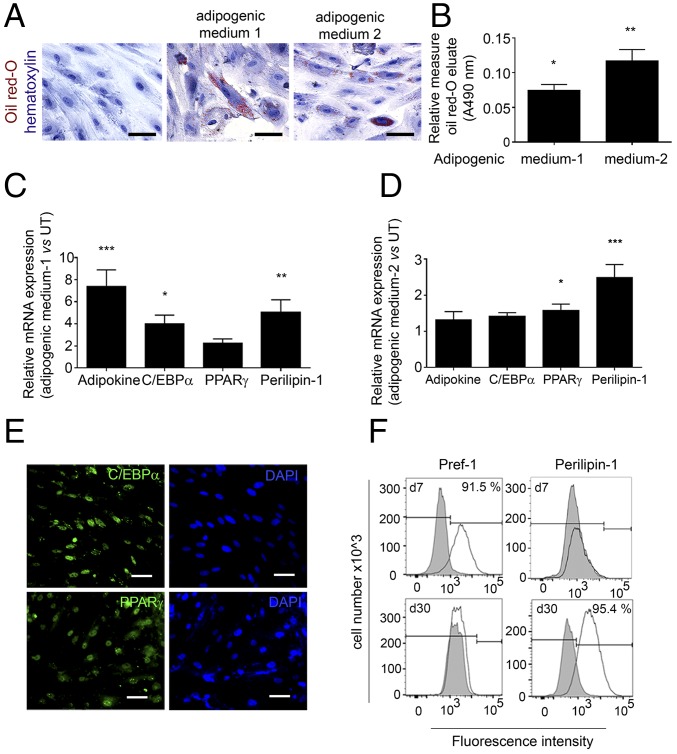
Fig. 2.
Human EPDCs have the capacity to differentiate into adipocytes in vitro. (A) Bright-field images of aEPDCs treated with adipogenic medium 1 or medium 2 or with basal medium stained with Oil Red O to identify lipid droplet formation and counterstained with hematoxylin (n = 35). (Scale bars, 20 µm.) (B) Histogram showing Oil Red O content (A490) (n = 35). Data represent values relative to untreated aEPDCs and are expressed as mean ± SEM of independent experiments, *P < 0.05, **P < 0.01, one-way ANOVA and Bonferroni’s post hoc test. (C and D) Expression of adipokine, C/EBPα, PPARγ, and Perilipin-1 in aEPDCs treated with adipogenic medium 1 (C) or medium 2 (D) (n = 10). Data represent the fold change relative to untreated aEPDCs and are expressed as the mean ± SEM of independent experiments. *P < 0.05, **P < 0.01, ***P < 0.001, one-way ANOVA and Bonferroni’s post hoc test. (E) Immunofluorescence staining for C/EBPα and PPARγ in aEPDCs treated with adipogenic medium 1. (Scale bars, 20 µm.) (F) Histograms show aEPDC Pref-1 and Perilipin-1 signal intensity analyzed by flow cytometry following treatment with adipogenic medium 2 for 7 or 30 d (n = 6). Isotype controls are shown in gray.

