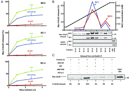
Fig. 1.
Isolation of an ATP-dependent disassembly factor from extracts of HeLa cells. (A) Part of the disassembly of MCC and of its subcomplexes by extracts of HeLa cells is ATP dependent, but phosphorylation independent. APC/C was removed from extracts of nocodazole-arrested HeLa cells by two sequential immunodepletions with anti-Cdc27, as described (10). Indicated volumes of this preparation were assayed for the disassembly of MCC and its subcomplexes, as described in Materials and Methods. Where indicated, the following additions were made: ATP, 2 mM; SP, 10 µM; and BI, 200 nM. Reactions without ATP were supplemented with hexokinase, 0.4 mg/mL and 2-deoxyglucose, 10 mM. (B) Anion exchange chromatography of ATP-dependent disassembly factor from extract of HeLa cells. Extracts from nocodazole-arrested HeLa cells (160 mg of protein) were applied to a 8-mL Source 15Q column (GE Healthcare) equilibrated with 50 mM Tris·HCl (pH 7.2), 50 mM NaCl, and 1 mM DTT. The column was washed with 30 mL of the above buffer and then subjected to a linear gradient of 50–450 mM NaCl (black squares) at a flow rate of 3.5 mL/min. Fractions of 8 mL were collected, concentrated by ultrafiltration, diluted 100-fold in a buffer consisting of 50 mM Tris·HCl (pH 7.2), 1 mM DTT, and 10% (vol/vol) glycerol, and concentrated again to a final volume of 1 mL. Samples of 1 μL of the indicated column fractions were assayed for the release of Cdc20 from MCC (red squares) and from BC-1 (blue squares) to the supernatant as described in Materials and Methods, in the presence of staurosporine and BI-2536. The Lower two panels are controls showing that BubR1 is not released from anti-BubR1 beads under the conditions of the disassembly activity assay, and therefore the release of Cdc20 is due to its dissociation from BubR1 in MCC. (C) The activity disassembly factor is dependent on ATP hydrolysis. Samples of 1 μL of anion exchange chromatography fraction 21 (B) were assayed for the release of Cdc20 from MCC bound to anti-BubR1 beads (Materials and Methods). Where indicated, additions were at the following concentrations: hexokinase (HK), 0.4 mg/mL; 2-deoxyglucose (2DG), 10 mM; ATP, AMP–PNP, and ATP-γ-S were supplemented at 2 mM.

