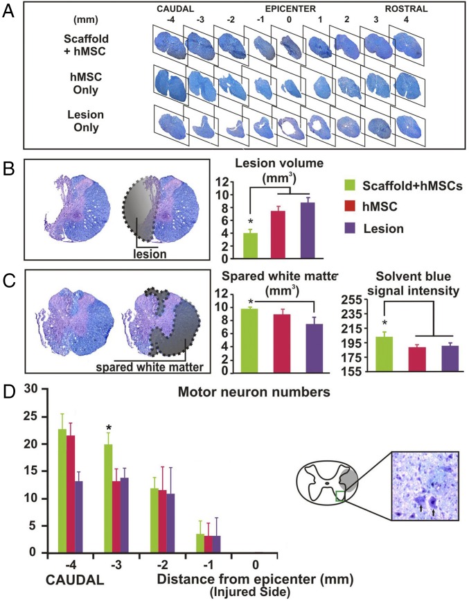
Fig. 3.
Histopathological analysis. Solvent blue- and hematoxylin-stained serial transverse spinal cord sections showed (A) that spinal cords with implanted scaffolded hMSC had more spared tissue around the lesion site than hMSC-alone and lesion-only controls and complete degradation of the PLGA scaffolds at ≥6 wk after SCI. Quantitative assessment of representative spinal cords (n = 4 per group) showed that relative to the controls, (B) scaffolded hMSC significantly reduced mean lesion volume and (C) increased white matter sparing in the spinal cord sections ±2 mm from the lesion epicenter. (D) Scaffolded hMSC implantation also preserved ventral horn motor neurons (Left) in spinal cord tissue caudal to the epicenter. Asterisks indicate a significant difference from the control group (P < 0.05, one-way ANOVA or repeated-measures ANOVA with Tukey’s post hoc test).

