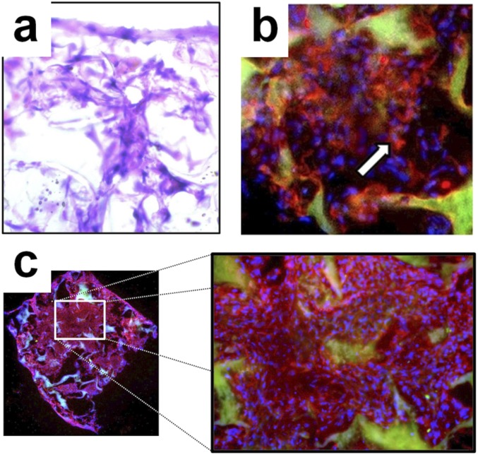
Fig. S2.
Characterization of scaffolded hMSCs. (A) H&E staining of hMSCs seeded in PLGA scaffold. The PLGA polymer scaffold has a porous, soft, and smooth texture (Materials and Methods). (B) Immunostaining revealed extensive BDNF expression in hMSCs seeded in PLGA scaffold (red, BDNF; blue, DAPI staining of cell nuclei; green, PLGA autofluorescence). (C) Immunocytochemical stain for CD90 showed a high level of hMSC engraftment in the tailored PLGA scaffold (red, CD90; blue, DAPI; green, PLGA autofluorescence).

