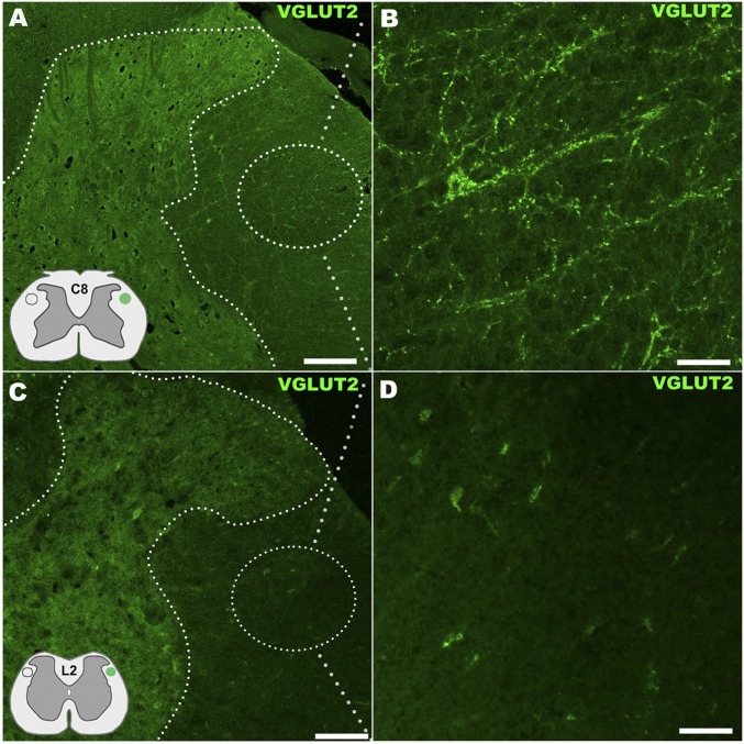
Fig. S4.
Assessment of RST innervation in the spinal cord by vGluT2 immunostain. (A) RST, located in the dorsal aspect of the lateral funiculus (Insets) showed innervation along the side of the spinal cord ipsilateral to and above T9–T10 hemisection in the scaffolded hMSC implantation group, with immunocytochemistry detecting vGluT2 that is carried by all RST terminals at C8 segment (area details in B). By contrast, no vGlut2 immunoreactive axon terminals or presence of RST neurites were detected in similar areas of spinal cord segments below the SCI site (C: at the L2 level with detail in D), indicating no regeneration of RST axons across the epicenter following scaffolded hMSC treatment. (Scale bars: A and C, 200 µm; B and D, 30 µm.)

