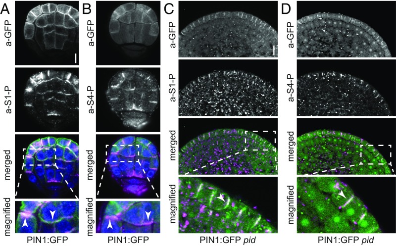
Fig. 6.
PIN1 phosphorylation follows PIN1 distribution in Arabidopsis embryos and pid shoot apical meristems. (A and B) Representative confocal images of Arabidopsis PIN1:GFP embryos stained with a-GFP (PIN1; green) and a-PIN1 S1-P (A; magenta) or a-PIN1 S4-P antibodies (B; magenta). Arrowheads mark the immunostaining at the plasma membrane. Merged images also include DAPI staining of nuclei (blue). (Scale bar, 5 µm.) (C and D) Representative confocal images of nondifferentiated pid PIN1:GFP mutant shoot apical meristems stained with a-GFP (PIN1; green) and a-PIN1 S1-P (C; magenta) or a-PIN1 S4-P antibodies (D; magenta). White arrowheads mark the immunostaining at the plasma membrane in the merged images. (Scale bar, 10 µm.)

