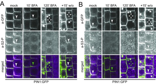
Fig. S1.
Phosphosite-specific a-PIN1 S2-P and S3-P antibodies detect PIN1 phosphorylation in a BFA-sensitive manner. Representative confocal images of stele cells of the primary root after immunostaining of 4-d-old PIN1:GFP Arabidopsis seedlings with anti-GFP (a-GFP, PIN1:GFP; green), a-PIN1 S2-P (magenta; A) and a-PIN1 S3-P (magenta; B) antibodies. Seedlings were mock-treated or BFA-treated for 10 and 120 min, respectively. A 15-min washout (w/o) followed the 120-min BFA treatment. Arrowheads mark strong and weak plasma membrane staining, asterisks mark intracellular compartments. Overlap of green and magenta signals is indicated by white arrows in the merged images. Green arrowheads and asterisks indicate the absence of a corresponding magenta signal from a-PIN1 S1-P or a-PIN1 S4-P immunostaining. (Scale bar, 5 µm.)

