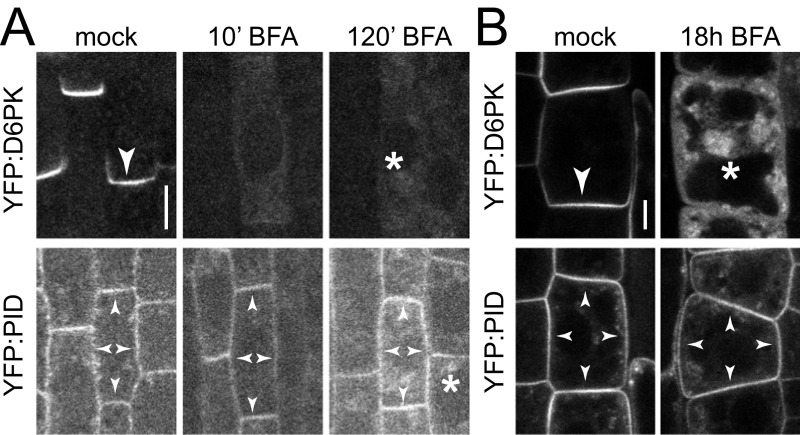
Fig. S3.
D6PK and PID have differential BFA-sensitivity in root stele and epidermis cells. Representative confocal images of (A) stele cells and (B) epidermal cells of the primary root from 4-d-old YFP:D6PK and YFP:PID Arabidopsis seedlings after mock or BFA treatments as used for the experiments shown in Figs. 1 and 5 and Fig. S1, followed by immunostaining (A) or live imaging (B). Arrowheads mark strong and weak plasma membrane staining, asterisks mark intracellular compartments. (Scale bars, 5 µm.)

