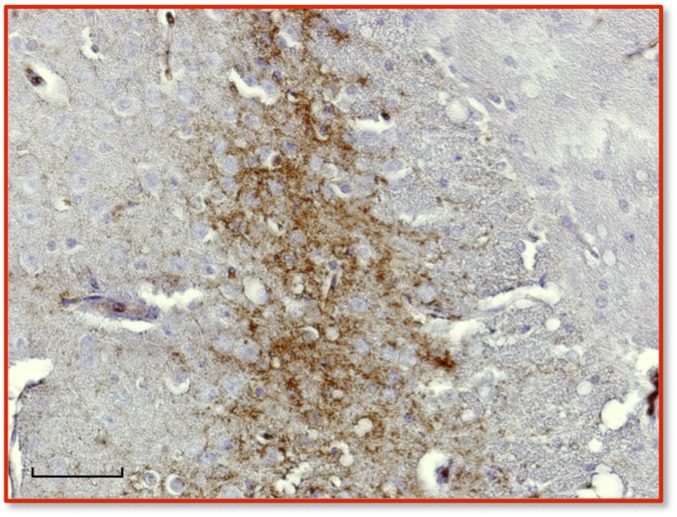
Fig. S2.
Immunohistochemical analysis of PrPSc in the central nervous system of diseased TgEq mice infected with SSBP/1. Immunohistochemical (IHC) analysis was performed as previously described (14) from sections of cortex taken at the region of the septum using mAb 6H4 as primary antibody and IgG1 biotinylated goat anti-mouse as secondary antibody (SouthernBiotech). Digitized images were obtained by light microscopy at 40× magnification using a Nikon Eclipse E600 microscope equipped with a Nikon DMX 1200F digital camera. (Scale bar, 50 μm.)

