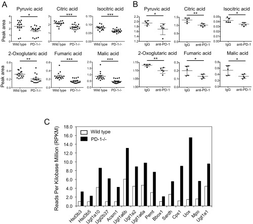
Fig. S1.
Mitochondrial activation in PD-1 blockade conditions. (A) The tricarboxylic acid cycle-associated metabolites in sera of wild-type and PD-1−/− mice were measured by gas-chromatography-mass spectrometry. The levels of the metabolites indicated were compared. Data represent the means ± SEM of 14 to 16 mice. *P < 0.05, **P < 0.01, ***P < 0.001, two-tailed Student t test. (B) MC38-bearing mice were treated with PD-1 mAb (4H2) according to the schedule described in Fig. 1A. TCA-associated metabolites were measured in sera harvested on day 13. *P < 0.05, **P < 0.01, two-tailed Student t test. (C) RNA extracted from lymph nodes of wild-type and PD-1−/− mice was subjected to RNA sequencing to compare the expression levels of genes associated with mitochondrial activity. The same amounts of RNA from seven individual mice were pooled. The mitochondria-related genes were selected using Gene Ontology GOterm_Cellular Component (CC)_FAT.

