Fig. 3.
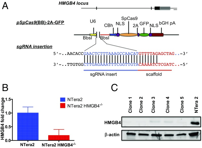
Targeting of the HMGB4 locus in mammalian embryonic carcinoma TGCT cells. (A) pSpCas9(BB)-2A-GFP expression vectors. The sgRNA cloned into the Bbs1 site of the Cas9-containing vector, encoding GFP for identification of transfected cells. GFP+ cells were sorted by FACS and single cells were expanded in culture. (B) mRNA levels of HMGB4 in parental NTera2 cells and knockout cells as determined by RT-PCR. (C) Western blot analysis of HMGB4 (Upper) protein expression 6 wk after pSpCas9(BB)-2A-GFP transfection. β-Actin (Lower) was used as loading control. Blots were probed with anti-HMGB4 or anti–β-actin antibody.

