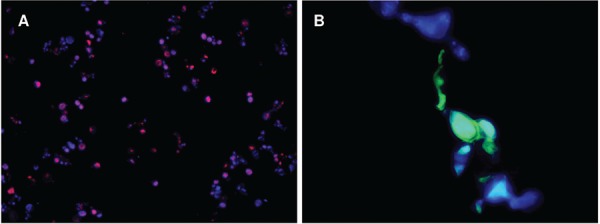
Fig. 3. : merged images of the yeast hybridisation with Paracoccidioides lutzii and P. brasiliensis specific probes. (A) Yeasts of P. lutzii hybridised with TEXAS-Red probe indicated by intracellular red dots (400x); (B) yeasts of P. brasiliensis hybridised with HRP-TSA probe indicated by intracellular green dots (1000x); (A, B) cellular walls can be visualised by structures marked in blue colour.

