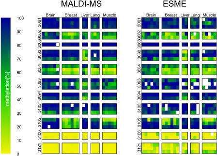
Figure 7. Comparison of Methylation Values Measured in Five Tissues and Eleven Amplicons Using MALDI-MS and ESME Analysis of Directly Sequenced PCR Products.
Each column is a tissue sample, each row a CpG site. Data are ordered in blocks by tissue type and amplicons. Positions of measurements for MALDI-MS (A) correspond to those for ESME analysis (B). The methylation values are colour coded from 0% methylation (yellow) to 100% methylation (blue), with intermediate methylation levels represented by shades of green. White indicates missing measurement values.

