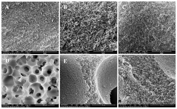
Figure 3.
SEM images of gelatin scaffolds. Both 2D and 3D GF scaffold morphology was observed by SEM after DFO conjugation. 2D GF at low magnification (A, scale bar = 50 μm) and high magnification (B, scale bar = 20 μm). 2D GF-DFO at high magnification (C, scale bar = 20 μm). 3D GF at low magnification (D, scale bar = 200 μm) and high magnification (E, scale bar = 20 μm). 3D GF-DFO scaffolds at high magnification (F, scale bar = 20 μm).

