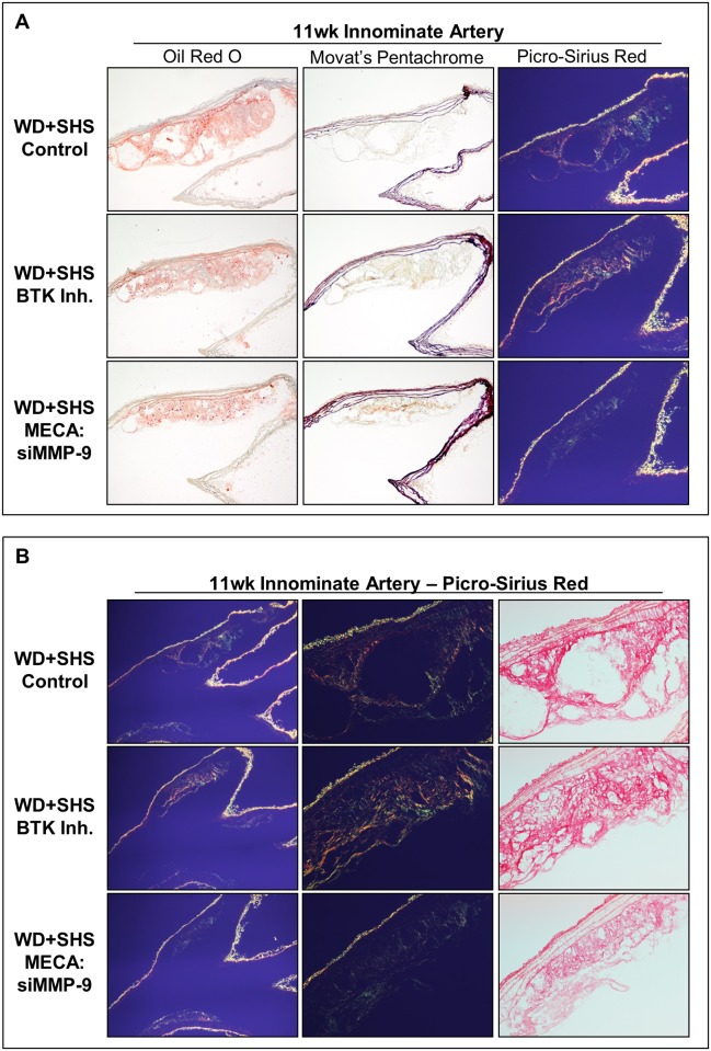
Fig 6. Histological analysis of aortic arch cross sections at the initial branch of the innominate artery from ApoE-/- mice following four week treatments.
(A) Representative Oil Red O stained sections of aortic arch show atherosclerotic lesions found at the initial branch of the innominate artery of control and treated mice; 4–8 animals per group were analyzed. Neutral lipids stained red. Movat’s Pentachrome and Picro-Sirius red stained sections of aortic arch are also shown; 3–5 mice per group were analyzed. (B) Representative Picro-Sirius red stained sections of aortic arch show atherosclerotic lesions found at the innominate artery of control and treated mice when viewed under normal (right) and polarized (left, center) light; 3–5 animals were evaluated per group.

