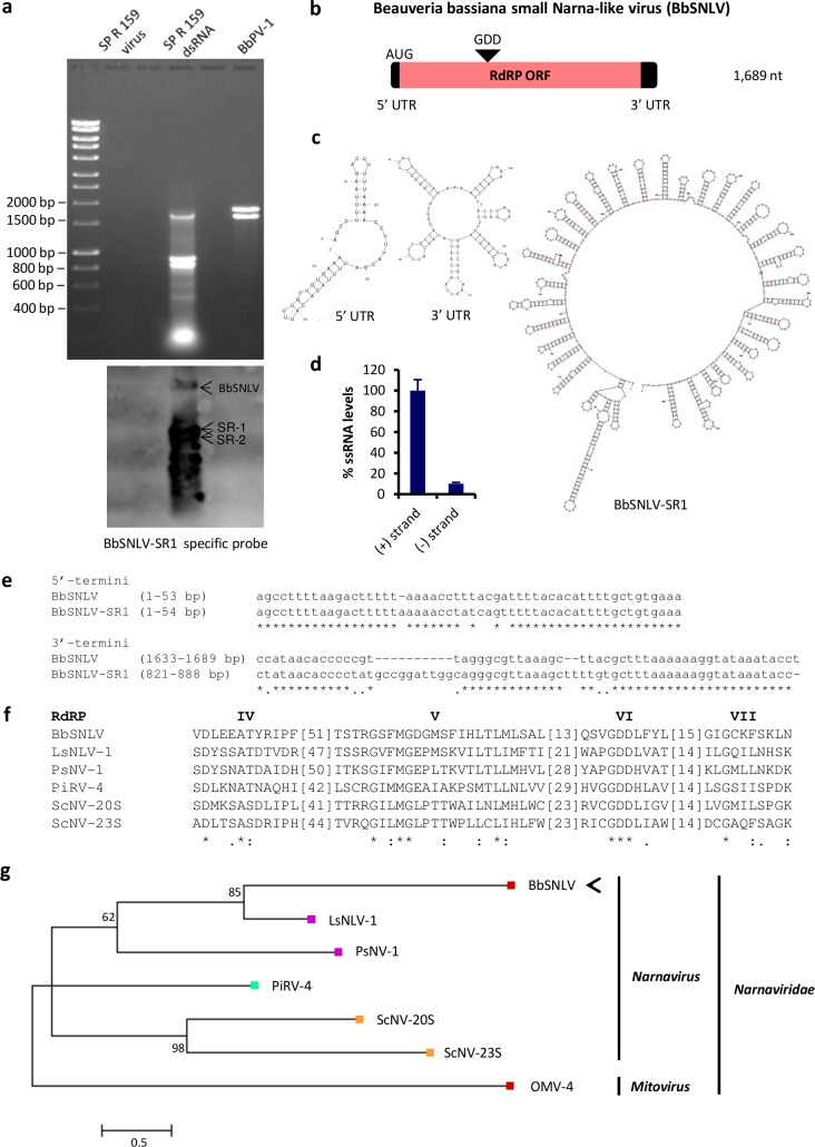
Fig 2. A small Narna-like virus in Beauveria bassiana.
(a) 1% (w/v) agarose gel electrophoresis and northern blot hybridization of dsRNA extracted from B. bassiana isolate SP R 159, using virus purification (lane 2) and LiCl dsRNA extraction (lane 4). Lane 6 contains partitivirus BbPV-1, as a negative control. Hybridization was carried out using specific probes for BbSNLV-SR-1: BbSNLV and SR 1 and 2 are indicated by arrows. (b) Schematic representation of the genomic organization of BbSNLV. The BbSNLV genome consists of a single RNA containing one ORF (grey box) flanked by 5’- and 3’-UTRs (black boxes). (c) Predicted secondary structure of the BbSNLV 5’- and 3’-UTRs and the (+) strand of BbSNLV-SR-1 using mfold (folding temperature 25°C). (d) Relative RNA levels of the positive and negative strands BbSNLV-SRs within total fungal ssRNA extracts, as shown by RT-qPCR amplification. At least three independent repetitions were performed in triplicate and error bars represent standard deviation. (e) A comparative alignment of the 5’- and 3’-terminal sequences of BbSNLV and BbSNLV-SR-1. Asterisks signify identical nucleotides. (f) A comparison of the conserved motifs of the RdRP in BbSNLV and other narnaviruses (S2 Table). Numbers within the brackets indicate the number of aa not shown. Asterisks signify identical aa residues, colons signify highly conserved residues and single dots signify less conserved but related residues. (g) Maximum likelihood phylogenetic tree created based on the alignment of RdRP sequences of narnaviruses (S2 Table) using the LG+G+I+F substitution model. Branches with bootstrap support lower that 50% were collapsed. At the end of the branches, red, orange, green and purple indicate that the virus infects filamentous fungi, yeast, oomycetes and kinetoplastids respectively. BbSNLV is indicated by an arrow.

