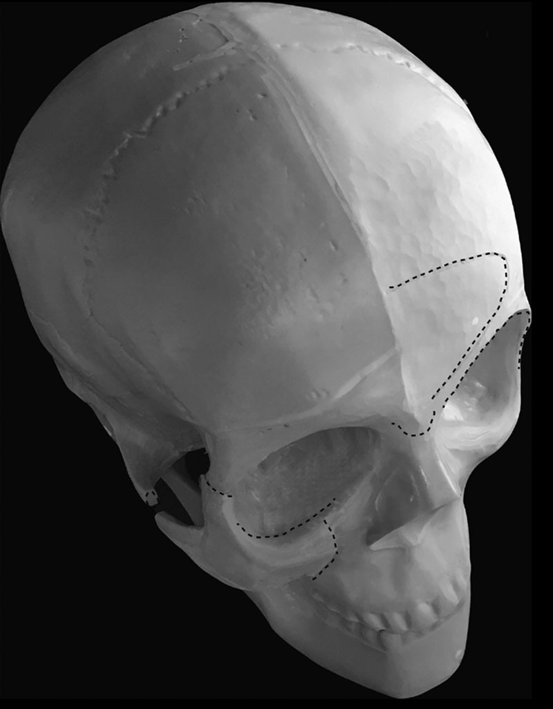
Fig. 1.

Model of the craniofacial skeleton. Right side, Midface osteotomies and anterior repositioning of the zygomatic/malar complex. Dotted lines denote areas of osteotomies. Left side, Dotted lines denote areas of upper face feminization of the frontal bone and orbit.
