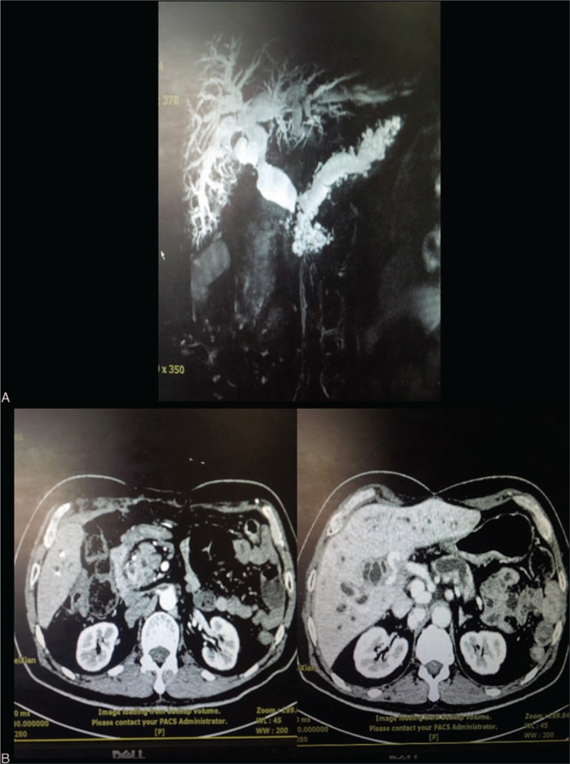
Figure 1.

(A) Magnetic resonance cholangiopancreatography (MRCP) reveals a mass in the ampulla of the duodenum of a 56-year-old male patient who developed Vater ampulla carcinoma 4 years after liver transplant surgery. (B) Abdomen enhancement CT demonstrates thickening of the lower segment of the common bile duct, and the main portal vein trunk and branches are not visualized. CT = computed tomography, MRCP = magnetic resonance cholangiopancreatography.
