
Figure 1.
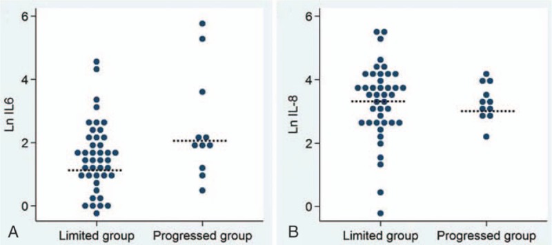
(A) The serum level of the Ln IL-6 in the limited and progressed group. The median serum level of Ln IL-6 of the progressed group was significantly greater than that of the limited group (2.0 vs 1.4, P = 0.025). (B) The serum level of the Ln IL-8 in the limited and progressed group. There was no significant correlation between the median serum level of Ln IL-8 and the tumor progression pattern (3.2 vs 3.5, P = 0.978). Ln = natural logarithm.