
Figure 1.
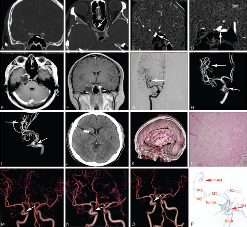
Brain CT and MRI examinations showed a hypointense lesion in the right parasellar and petrous apex region (A–F). DSA and 3D-DSA images demonstrated an right saccular aneurysm (arrow) originated from the M2–M3 junction of the right MCA and a saccular aneurysm of the clinoid segment of right internal carotid artery (ICA) (G–I). Postoperative CT and 3D-CTA images (J). The diagnosis of epidermoid tumor was confirmed by pathologic examination (L). 3D-CTA images showed the morphology of the untreated aneurysm of the clinoid segment of right ICA remained stable 3 times in 5-years follow-up (M–O). Illustration demonstrating the relative position between the epidermoid tumor and the untreated aneurysm (P).