Figure 1.

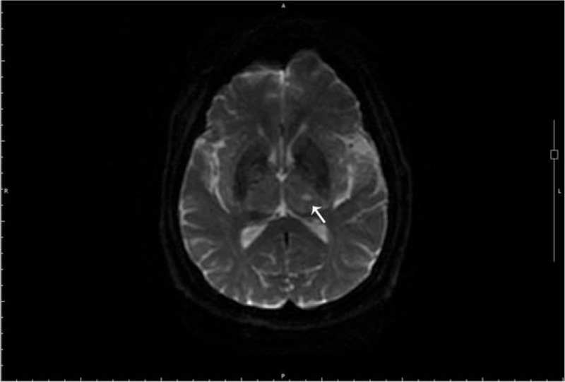
Brain MRI-DWI shows an old infarction focus in the left thalamus (white arrow). MRI-DWI = magnetic resonance image diffusion weighted imaging.

Brain MRI-DWI shows an old infarction focus in the left thalamus (white arrow). MRI-DWI = magnetic resonance image diffusion weighted imaging.