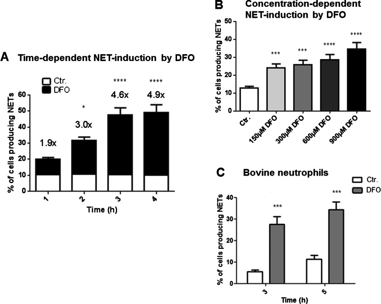
Figure 4. DFO-induced NET formation is time- and concentration dependent and not limited to human neutrophils.
Human and bovine blood-derived neutrophils were isolated by density gradient centrifugation, stimulated and the formation of NETs was visualized using the PL2–6 antibody against H2A–H2B–DNA complexes (green) in combination with DAPI to stain the nuclei (blue). (A) Human neutrophils were stimulated with 300 μM DFO for 1, 2, 3 and 4 h and subsequently fixed in 4% PFA. NET formation was determined in comparison with the unstimulated control. A significant increase in the amount of cells that form NETs was observed over time. The numbers on top of the bars represent the fold increase in NET-release from cells treated with DFO compared with the unstimulated control. (B) Different DFO concentrations (150, 300, 600, 900 μM) were tested on their ability to induce NETs in human neutrophils after an incubation period of 3 h. (C) NET formation in bovine neutrophils after stimulation with either media only or media containing 300 μM DFO for 3 and 5 h. The graphs represent the mean ± S.E.M. of the 24 (A), 30 (B), 12 (C) images derived from 4 (A), 5 (B), 2 (C) independent experiments.

