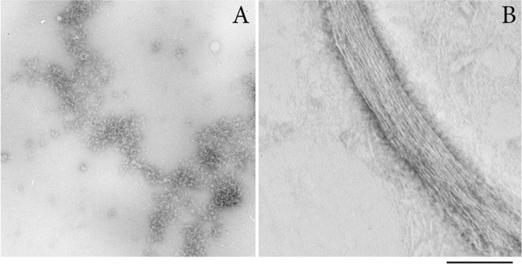
Figure 2. EM of negatively stained SMT aggregates.
(A) Amorphous titin aggregates are the main form of the aggregated protein. (B) A bundle of linear fibrils. The width of one fibril in the bundle is approximately 2 nm. SMT aggregates were obtained by 24 h dialysis in 0.15 M glycine–KOH, pH 7.0, at 4°C. Staining used 2% aqueous uranyl acetate, scale 100 nm.

