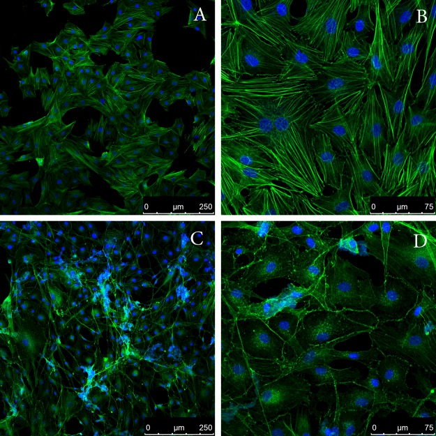
Figure 8. Addition of SMT amyloid to the culture media leads to disorganization of the actin cytoskeleton of aortic smooth muscle cells.
Panels show confocal microscopy of bovine aortic smooth muscle cells stained with phalloidin Atto 488. (A and B) Smooth muscle cells in the presence of actin fibrils (control); (C and D) smooth muscle cells in the presence of SMT amyloid aggregates. Incubation time was 48 h.

