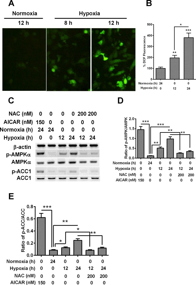
Figure 3. AMPK is activated by hypoxia via oxidant signalling.
(A and B) DCFDA detection for cellular ROS in H9C2 cells under normoxia or hypoxia, the representive image of DCF staining (A) and the quantitative analysis (B) was in indicated. (C) Western blotting of AMPK signalling in H9C2 cells under hypoxia and treated with AICAR or NAC. (D and E) Levels of AMPK phosphorylated at Thr172 (p-AMPK) (D) and of ACC phosphorylated at Ser79 (p-ACC) (E) respectively relative to AMPK and ACC, as was presented as fold change in the p-AMPK/AMPK or p-ACC/ACC ratio; *P<0.05, **P<0.01, ***P<0.001.

