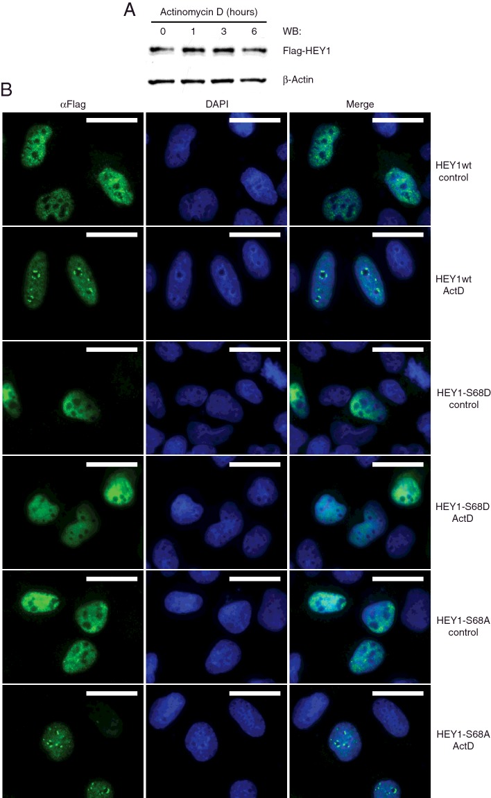
Figure 11. Ribosomal stress induced by actinomycin D causes HEY1 perinucleolar localization.
(A) U2OS cells previously transfected with expression vector for Flag-tagged HEY1 were treated with actinomycin D (5 nM) for 1, 3 or 6 h. HEY1 total protein levels were analysed by western blotting using anti-Flag antibody. (B) U2OS cells previously transfected with expression vectors for Flag-tagged HEY1, HEY1-S68D or HEY1-S68A were treated with actinomycin D (5 nM) for 6 h and assayed by indirect immunofluorescence with anti-Flag antibody. The first column shows the indirect immunofluorescence with anti-Flag antibody, the second column shows DAPI staining of DNA and the third column shows the merge image indicating the degree of colocalization. Bars, 20 μm.

