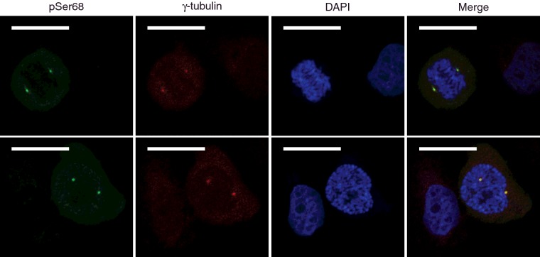
Figure 8. Confocal section of U2OS-HEY1 cells showing high concentration of HEY1 phosphorylated at Ser-68 in the centrosomes of mitotic cells.
U2OS-HEY1 cells were treated with 1 μg/ml tetracycline to induce expression of HEY1. After 6 h cells were fixed and processed for double immunofluorescence staining using antibodies specific for HEY1-phospho-S68 and the centrosome marker γ-tubulin. DAPI is used to counterstain the nucleus. Bars, 20 μm. Two representative confocal microscopy images are shown.

