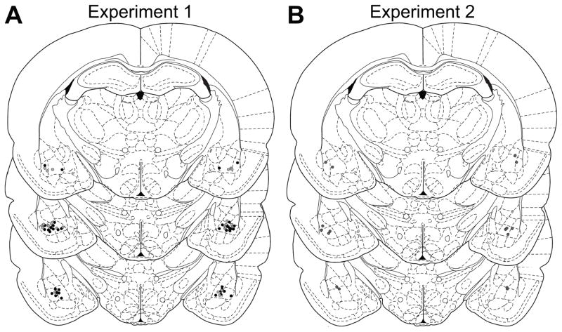
Figure 1. Histological verification of BLA cannula placements.
Schematic representation of microinfusion injector tips for Experiment 1 (A; black, delta group; gray, mu group) or Experiment 2 (B). Line drawings of each section taken from (Paxinos & Watson, 1998) −2.8 – 3.3 mm posterior from bregma.

