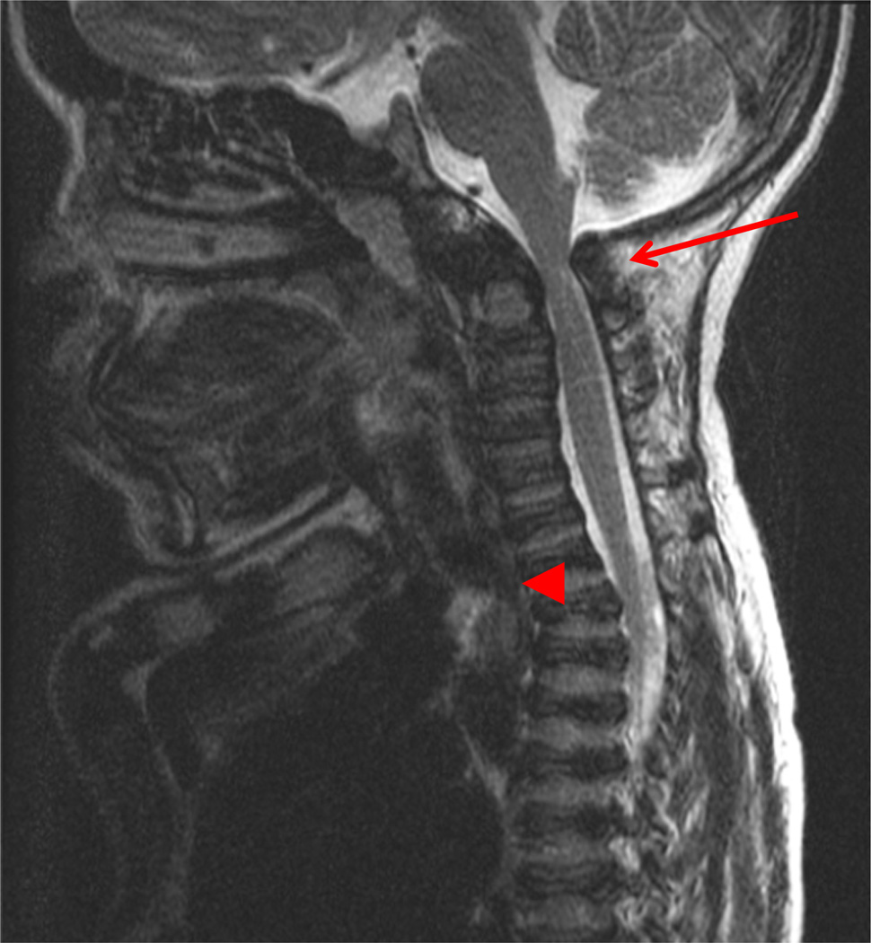
Fig. 3.
MRI of cervical spine in a 13 years patient. The arrow shows C1-C2 spinal cord compression and the arrow head specifies tracheal obstruction. A baseline study of the upper cervical anatomy is recommended no later than 2 years or at diagnosis using flexion/extension X-ray films. If severe pain or pain associated with weakness of strength or tremors (or clonus) in the arms or legs occur, the patient should have studies of the neck to evaluate for the slippage (subluxation) of the neck vertebrae and compression of the spinal cord.

