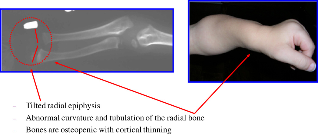
Fig. 4.
Characteristic bone deformity in the upper extremity (adapted from Educational CD for Morquio and permitted by Carol Ann Foundation). The epiphyseal involvement characteristic of MPS IVA is exemplified by the tapered irregular distal radius and ulna. The bones are osteopenic with cortical thinning. Upper extremities in a child aged 2 years 3 months (left panel). Note the irregular epiphyses and widened metaphyses. Cortical thinning and mild widening of the diaphysis of the humerus are visible. With age, the bone deformity progresses, e.g. with tilting of the radial epiphysis towards the ulna (10 years old; right panel). The humerus usually appears shortened later.

