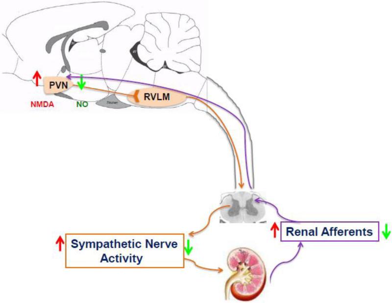
Figure 5.
Schematic diagram illustrating that sensory information originating in the kidney is transmitted by renal afferent nerves to the dorsal column of the spinal cord which results in exciting pre-autonomic neurons in the PVN via multiple synaptic pathway. The pre-autonomic neurons in the PVN project to the RVLM as well as the intermediary column of the spinal cord, where pre-sympathetic neurons terminating in the kidney reside. These pre-autonomic neurons are activated by NMDA and inhibited by NO. This long loop pathway involving the PVN suggests that this renal-PVN pathway may contribute to the elevated sympathetic nerve activity in disease conditions such as CHF. Renal denervation reduces afferent renal nerve signaling, neuronal activity in the PVN and sympathetic nerve activity in the CHF. RVLM, rostral ventrolateral medulla; NMDA, N-methyl-d-aspartate; NO, nitric oxide. Orange arrows indicate efferent pathway affecting the kidney. Purple arrows indicate afferent pathway to the PVN. Red arrows: indicate changes during CHF; green arrows: indicate changes after renal denervation.

