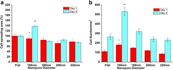
Fig. 3.

Statistical analysis of cell spreading area (%) and cell number (/mm2) on different nanoporous stainless steel artificial microenvironments. a After day 1, no significant difference in the cell spreading area was observed in cells on control and 100-nm nanoporous surfaces. After day 3, cell spreading area percentage maximized at 100 nm. Consistent decrement in cell spreading area percentage was observed as the nanopore diameter became more than 100 nm. One hundred-nanometer nanoporous surfaces acted as the transition-inducing factor for cell spreading area. b After day 1, cell number maximized at 100-nm nanoporous surfaces. Consistent decrement in cell number was observed as the nanopore diameter became more than 100 nm. One hundred-nanometer nanopore diameter acted as the transition-inducing factor for cell number. Statistically significant data is represented by * having p < 0.05; highly significant values are represented with ** having p < 0.01
