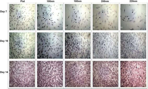
Fig. 7.

Alizarin red S staining to visualize mineral calcium deposition. Cells were cultured on different nanoporous artificial microenvironments for 7, 10, and 14 days, and alizarin red S staining was then performed. Maximum staining intensity signifying maximum calcium mineral deposition was observed in cells on 100-nm nanoporous surfaces. Arrows point to the purple-stained sections on nanosurfaces. Mineral deposition maximized on day 14 in cells on 100-nm nanoporous surfaces. Scale bar represents 25 μm
