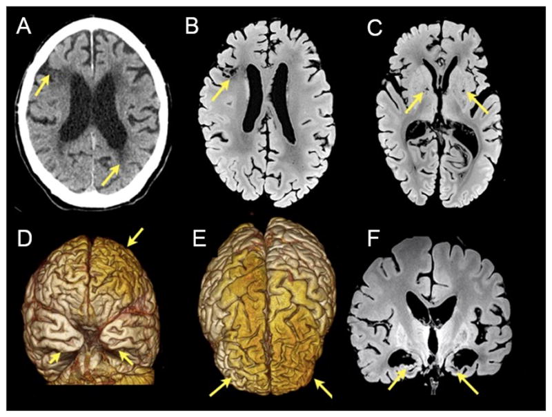
Figure 1.

Radiological features of the case. A) Non-contrast axial CT: the arrow points to hypoattenuation in periventricular white matter region and infarct in the right middle frontal gyrus (arrow). B) and C) Axial formalin fixed post-mortem MR T1-weighted images showing the infarct (arrow in B) and lacunae in the basal ganglia (arrows in C). D) Frontal and E) superior views from 3D renderings from the postmortem MR images showing atrophic changes in frontal and temporal areas (arrows). F) Coronal formalin fixed post-mortem MR T1-weighted images showing a marked reduction of the mesial temporal and hypothalamic structures.
