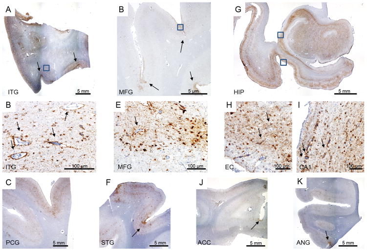
Figure 3.
CTE pathology. A) Inferior temporal gyrus (ITG) depicting widespread tau pathology, more concentrated in the superficial cortical layers and depth of the sulci (arrows). B) Enlargement of the boxed area on (A) highlighting the predominately glial pathology and perivascular tau pathology (arrows). C) Widespread tau pathology in precentral gyrus (PCG). D) Middle frontal gyrus showing tau pathology especially at the depth of the sulci (arrow). E) Enlargement of the boxed area on (D) depicting tau glial and neuronal pathology and perivascular tau deposits (arrow). F) Tau pathology in superior temporal gyrus (STG), more prominent in the depths of the sulcus (arrow). G) Hippocampal formation showing widespread tau pathology. Using this staining, it is possible to observe the thinning of the CA1 sector and subiculum. H) Enlargement over the entorhinal cortex (bottom box on G) again showing a predominant glial tau pathology and perivascular tau deposits (arrow). I) Enlargement over the CA1 sector (top box on G) highlighting the perivascular tau pathology (arrow). J and I) Tau pathology more prominent in the depth of the sulcus (arrows) in anterior cingulate cortex (ACC) and angular gyrus (ANG), respectively. All histological sections were immunostained for phospho-tau (CP13, 1:500, gift of Peter Davies).

