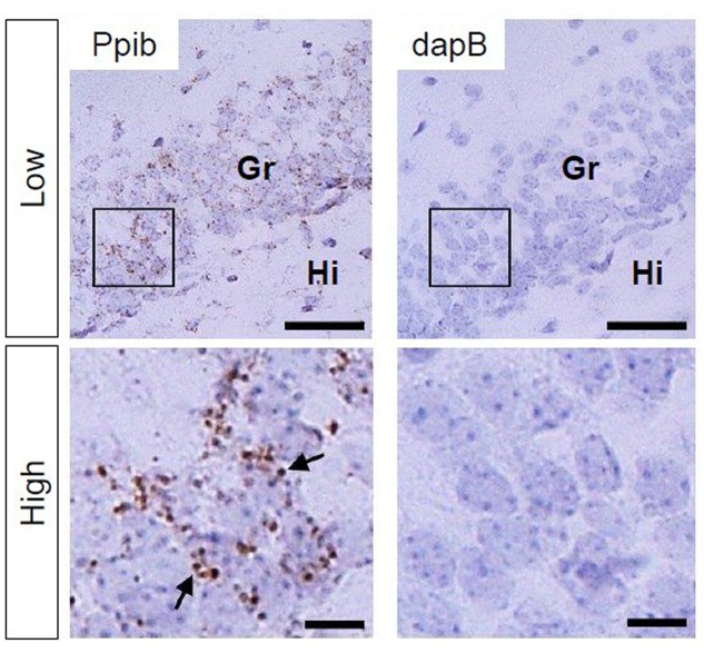
FIGURE 2.
Peptidylprolyl isomerase (Ppib) and dapB mRNA expression in the hippocampal DG in PND 14 mice. Brown punctae (arrows) represent Ppib mRNA (left). No signal associated with dapB mRNA was observed (right). “Low” and “High” indicate magnification. High magnification images represent the areas enclosed by boxes in the low-magnification images. Scale bars = 50 and 10 μm in low and high magnification images, respectively. Gr, granule cell layer; Hi, hilus.

