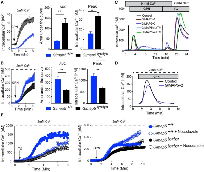
Figure 7.
GPN-mediated release of Ca2+ is increased in the absence of GIMAP5. Peripheral CD4+ T lymphocytes purified from Gimap5lyp/lyp and control rats were loaded with Fura-2 and intracellular Ca2+ was measured as described. The arrow indicates the time of addition of GPN (100 µM). Cytosolic Ca2+ was measured following addition of GPN in the absence (A) or presence (B) of extracellular Ca2+. The histograms represent the area under curve (AUC) and the peak values. Student’s t-test (**p < 0.005). (C) Cytosolic Ca2+ was measured using Fura-2-labeled stable transfectants of the indicated GIMAP5 constructs. The cells were maintained in Ca2+-free medium (0 mM Ca2+ and 0.5 mM EGTA), and GPN and TG were added sequentially as indicated before adding Ca2+ to the extracellular medium. (D) Ca2+ release from stable transfectants of the vector control or GIMAP5v2 following addition of GPN in the presence of extracellular Ca2+. (E) Cytosolic Ca2+ was measured following addition of TG or GPN in the presence of extracellular Ca2+ in T lymphocytes pretreated or not with nocodazole (2 µM) for 30 min.

