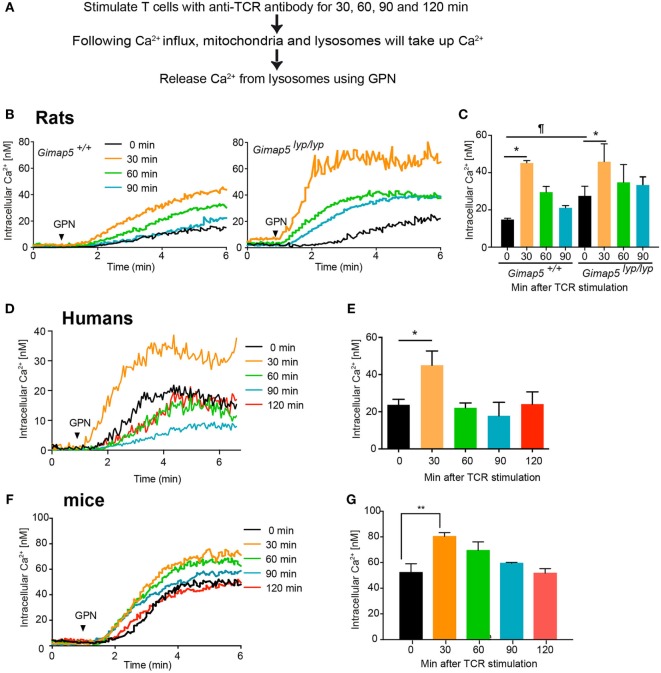
Figure 8.
Lysosomes accumulate Ca2+ following activation through T cell receptor. (A) Schematic representation of the experimental setup is indicated. Purified CD4+ T lymphocytes from (B,C) rats, (D,E) humans, and (F,G) mice were plated on coverslips and stimulated with anti-TCR antibody (R73 for rats, IP26 for humans, and H57 for mice) for the indicated duration. The lymphocytes were then loaded with Fura-2. Lysosomal Ca2+ content was probed with GPN in the absence of extracellular Ca2+. Representative data from one experiment are shown in (B,D,F). The average of peak values from three independent experiments is shown in (C,E,G). ¶p < 0.05 between WT and Gimap5lyp/lyp unstimulated (*p < 0.05; **p < 0.01).

