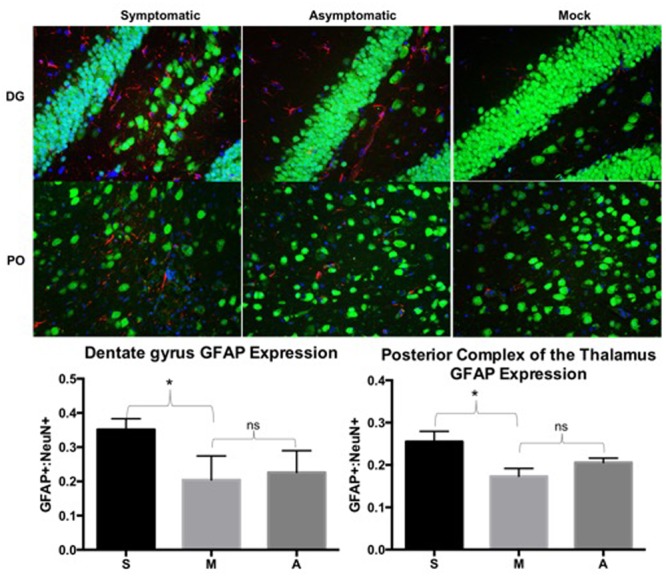
FIGURE 4.

Symptomatic animals had more GFAP expression than both asymptomatic and mock groups in the dentate gyrus (DG) and posterior complex of the thalamus (PO). Asymptomatic animals had more GFAP expression than the mock animals, but less so than symptomatic animals. GFAP expression was determined by dividing counts of GFAP positive cells by NeuN positive cells in comparable fields of view. Representative images from each group are depicted. Green: NeuN, Red: GFAP, Blue: DAPI. ns: not significant, ∗p < 0.005. S: symptomatic; M: mock; A: asymptomatic.
