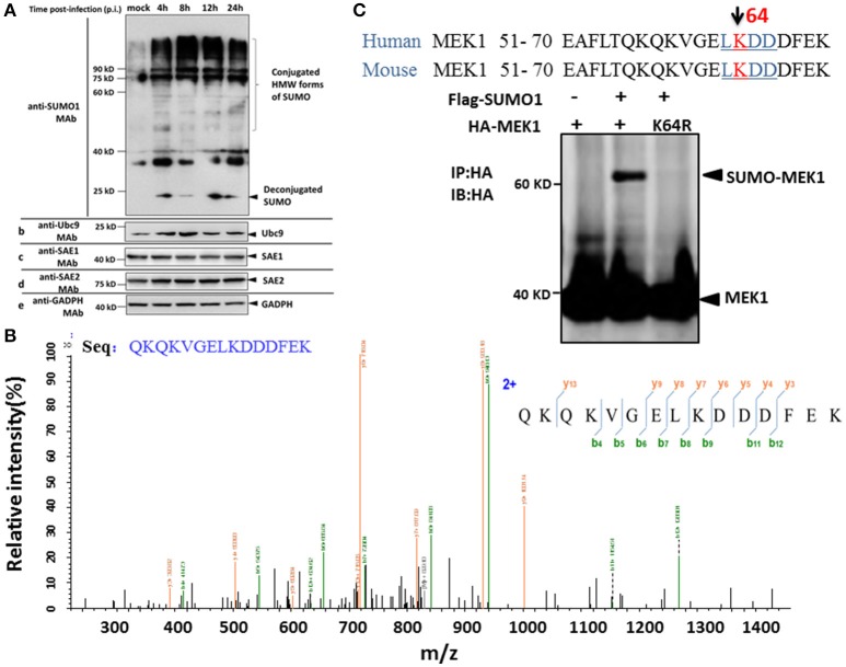
Figure 1.
SUMOylation of MEK1 is decreased in A549 cells after H5N1 influenza virus infection. (A) A549 cells were infected with H5N1 at an MOI of 7, total cell extracts were collected at 4, 8, 12, and 24 h post infection, and analyzed by SDS-PAGE and western blotting using MAbs to SUMO1, Ubc9, SAE1, SAE2, and an internal control GAPDH. (B) Identification of the MEK1 SUMOylation site by LC-MS/MS analysis. (C) Alignment of MEK1 N-terminal amino acids from human and mouse. Bold and underlined text highlights the conserved LKDD motif.

