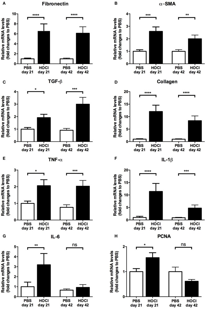
Figure 2.
Expression of fibrosis, inflammation, and proliferation markers in the skin of phosphate-buffered saline (PBS) and HOCl mice at day 21 and day 42 of the protocol. mRNA levels of various markers of fibrosis [fibronectin (A), α-smooth muscle actin (α-SMA) (B), transforming growth factor (TGF)-β (C), collagen (D)], inflammation [tumor necrosis factor (TNF)-α (E), interleukin (IL)-1β (F), IL-6 (G)], and proliferation [proliferating cell nuclear antigen (PCNA) (H)], normalized to GAPDH and expressed as fold changes to PBS day 21 (for day-21 groups) or to PBS day 42 (for day-42 groups) (n = 15–24 per group).

