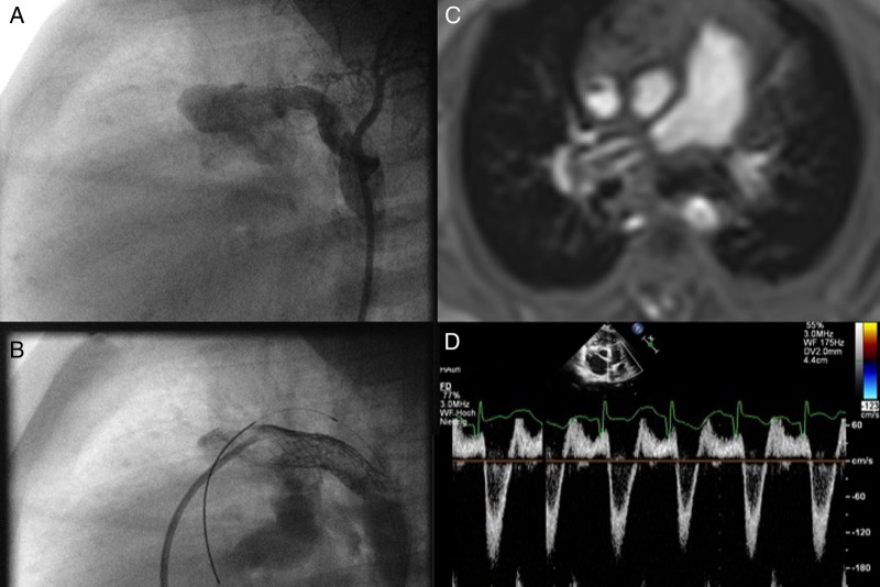
Figure 4.
(A–D) The need for a valved Potts shunt. (A) shows a surgically performed reversed ‘Potts’ shunt with a dimension of 6 mm; in the follow-up, the polytetrafluorethylen (PTFE)-shunt was dilated by placing a 8 mm Formula Stent (B); but despite a bilateral pulmonary banding, which is depicted on MRI (C), the Doppler echocardiography (D) shows a systolic right-to-left, but a diastolic left-to-right shunt flow.

