
Figure 3.
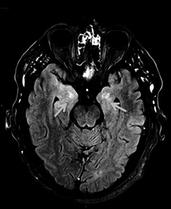
MRI brain postcontrast FLAIR imaging showing asymmetric bilateral abnormal high signal in the mesial temporal lobes with abnormal parenchymal enhancement. FLAIR, fluid-attenuated inversion recovery.
Official websites use .gov
A
.gov website belongs to an official
government organization in the United States.
Secure .gov websites use HTTPS
A lock (
) or https:// means you've safely
connected to the .gov website. Share sensitive
information only on official, secure websites.
MRI brain postcontrast FLAIR imaging showing asymmetric bilateral abnormal high signal in the mesial temporal lobes with abnormal parenchymal enhancement. FLAIR, fluid-attenuated inversion recovery.