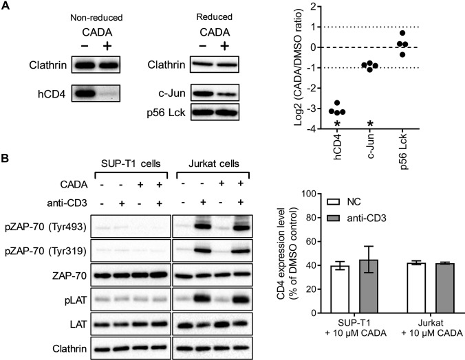
Fig. 4.
CADA down-modulates hCD4 and c-Jun but does not affect phosphorylation events involved in early TCR signaling. (A) DMSO and CADA-treated (10 μm, 72 h) SUP-T1 samples were blotted under nonreducing (left) and reducing conditions (right). Selected proteins were identified with conditions optimized for each individual primary antibody and representative images are shown for each protein. Quantification was performed as in Fig. 3A. (B) SUP-T1 and Jurkat cells were grown in the presence of DMSO or CADA (10 μm) for 24 h. Phosphorylation of TCR signaling proteins was then induced through cross-linking of CD3, followed by cell lysis and Western blotting (left panel; one representative set of blots out of two experiments is shown). Flow cytometric quantification of surface hCD4 levels on these cells shows that the down-modulation of hCD4 by 24 h CADA-treatment was comparable between SUP-T1 and Jurkat cells, independent of activation by crosslinking CD3 (right panel; bars are mean ± S.E., n = 2).

