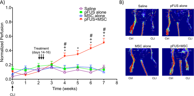
Figure 4. Weekly laser Doppler perfusion imaging (LDPI) in the feet of mice.
(A) Mice had critical limb ischemia (CLI) induced on Week 0. Two weeks post-surgery, mice were treated for 3 consecutive days (Day 14, 15, and 16) with: saline (n = 8), pFUS (n = 7), mesenchymal stem cells (MSC 106/day IV, n = 8) and pulsed focused ultrasound (pFUS) + MSC (106/day IV, n = 17) and imaged by LDPI weekly for another 5 weeks (7 weeks post-surgery). Statistical significance (p < 0.05; two-way ANOVA) of the pFUS + MSC groups is indicated by * compared to the saline group, + compared to the MSC only group, # compared to the pFUS only group. (B) Representative LDPI of saline, pFUS, MSC, and pFUS + MSC at 7 weeks post induction of CLI.

