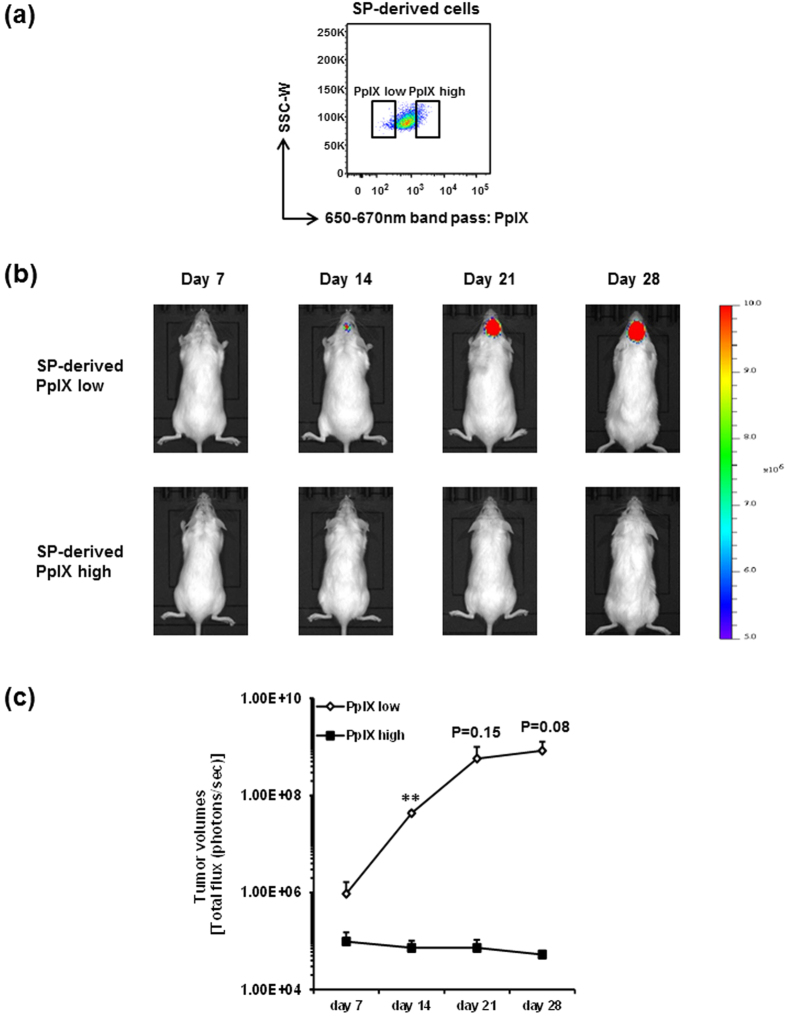
Figure 3. C6 SP-derived PpIX fluorescence low cells possess the tumorigenic capacity.
(a) Representative FACS plot of PpIX fluorescence low and high cells in C6 SP-derived cells treated with 5-ALA. SP-derived PpIX fluorescence low and high cells were sorted and intracranially transplanted into NOD/SCID mice. (b) Representative IVIS images of tumor acquired at day 7, 14, 21 and 28 after transplantation were displayed. Images from other two series of experiments are shown in Supplementary Figure 1. All IVIS images are presented at the same min-max threshold. (c) Tumor volumes are assessed by total flux of luminescence signals and shown as means ± SD from three independent experiments. **P < 0.01.

