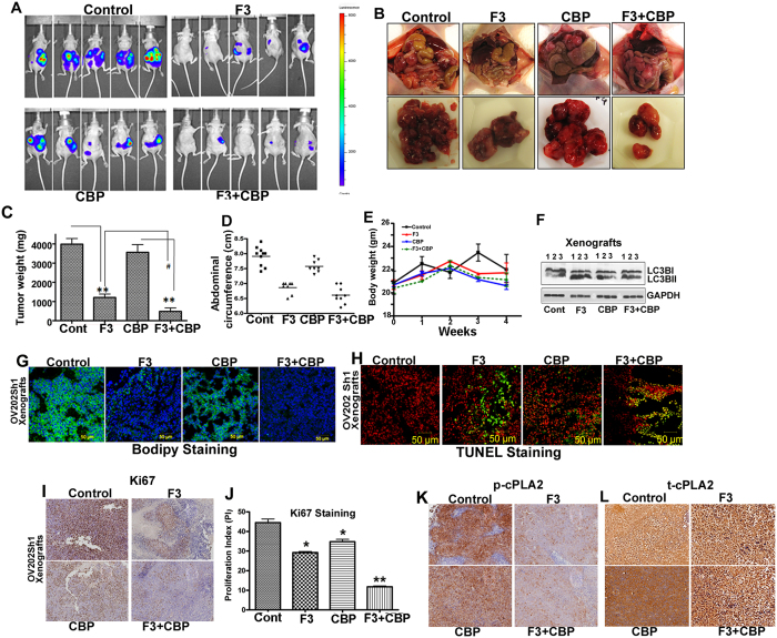
Figure 5. AACOCF3 alone and in combination with carboplatin suppresses tumor growth, and inhibits lipid droplet biogenesis in vivo.
(A) Randomized OV202 Sh1 tumor-bearing mice (10 mice per group), were treated with water, AACOCF3 (F3) (10 mg/kg), carboplatin (CBP) (51 mg/kg) or with a combination of AACOCF3 and CBP for 4 weeks as detailed in the manuscript. Mice were euthanized after 28 days. Representative images of the mice prior to sacrifice from control, F3, CBP, and CBP + F3 treatment groups using IVIS luminescence imaging system series 2000 are presented. Color barn indicates photon intensity. (B) Representative images of excised tumors from OV202 Sh1 xenografts. (C) Excised tumor weights from control, F3, CBP, and CBP + F3 treatment groups. (D) Abdominal circumference of the mice measured on the day of sacrifice across treatment groups. (E) Total body weight of control, F3, CBP, and CBP + F3 treatment groups.(F) Immunoblot analysis of LC3BI and LC3BII in lysates from control, F3, CBP, and F3 + CBP treatment groups. GAPDH was used as loading control. (G) Tumor xenografts were stained with Bodipy stain to detect lipid droplets (LD). DAPI was used to stain the nuclei. (H) Representative images of TUNEL staining of frozen section of xenografts from control, F3, CBP, and CBP + F3 treatment groups. Green fluorescence indicates TUNEL and red fluorescence indicates propidium iodide staining. (I) Representative images of immunohistochemical staining of Ki67. (J) Bar graph showing the Ki67 proliferation index (*P < 0.05 and **P < 0.01). The Ki67 proliferation index was measured using ImmunoRatio (public domain image analysis software). (K) p-cPLA2 in formalin-fixed, paraffin-embedded sections of treated and untreated xenografts.

