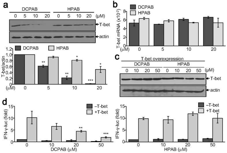
Figure 4. Decreased T-bet expression by treatment with DCPAB and HPAB.
(a,b) CD4+ T cells were treated with DCPAB or HPAB for 48 h under Th1-skewing conditions. Proteins were extracted and resolved by SDS-PAGE. Protein blots were incubated with anti-T-bet Ab, followed by detection using ECL and densitometry analysis (a). *P < 0.05, **P < 0.005, ***P < 0.0005. Total RNA was independently prepared using TRIzol reagent and subjected to reverse transcription, followed by the quantitative analysis of T-bet mRNA (b). (c,d) HEK 293 T cells were transfected with T-bet expression vector together with INF-γ promoter-linked reporter gene (pIFN-γ-luc). The β-galactosidase expression vector, pCMVβ was also transfected for normalizing transfection efficiency. Transfected cells were incubated with DCPAB or HPAB for an additional 24 h. T-bet expression was determined by immunoblot analysis (c). Relative luciferase activity was calculated after normalization with β-galactosidase activity and is expressed as a fold induction from four independent experiments (d). **P < 0.005, ***P < 0.0005.

