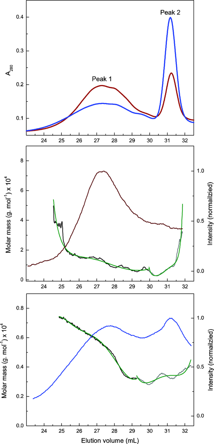
Figure 4. GPC-MALS analysis of APPL extracted from SPP incubated without (brown) or with sLac (blue).

The top panel shows the response from the multi-wavelength detector (280 nm). The lower panels show the response of the MALS detector to APPL isolated from SPP without (middle) and with (bottom) sLac. The green curves represent multiple exponential fits of the molar mass distribution to the MALS data obtained from the areas under Peaks 1 (black) and 2 (grey).
