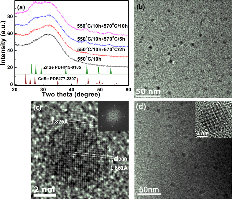
Figure 5.
(a) X-ray diffraction patterns of C4-0, C4-2, C4-5 and C4-10 specimens, (b) TEM image of C4-10 specimen and (c) one nanocrystal formed in C4-10 specimen, (d) TEM image of C4-0 specimen. The inset in (c) is the fast Fourier transformation image of the nanocrystal shown in (c), and the inset in (d) shows the HR-TEM image of one nanocrystal formed in C4-0 specimen.

